

Marcelo C. Ventura Filho1; Liana O. Ventura2; Camila V. Ventura3; Rubens Belfort Jr.4; Mauricio Maia5
DOI: 10.17545/eoftalmo/2018.0001
ABSTRACT
The emergence of Zika virus (ZIKV), which was previously limited to sporadic cases in Africa and Asia, with subclinical or mild influenza-like illness, has rapidly become widespread in the Americas since 2015, exhibiting a relationship with microcephaly and other birth defects. The broad spectrum of adverse outcomes caused by ZIKV infection in utero has been described as congenital Zika syndrome (CZS). One of the important pillars of this new entity is the ocular manifestations that may be presented at birth by the affected newborns. Interestingly, all infants with CZS show significant visual impairment, regardless of whether they present with ocular findings, which suggests cortical visual impairment. In addition, infants with CZS may have limited power of accommodation, poor visual acuity, high refractive errors, and strabismus, which frequently require refractive correction. Thus, CZS is a challenge not only to families but also to health care providers and the public health system.
Keywords: Zika Virus; Eye Manifestations; Visual Impairment.
RESUMO
O vírus da zika (ZIKV), que anteriormente se limitava a casos esporádicos na África e na Ásia, com sintomas similares a uma gripe leve ou subclínica, rapidamente tomou conta das Américas desde 2015 e passou a ter comprovadamente uma relação com microcefalia e outros defeitos congênitos. O amplo espectro das consequências causadas pela infecção intrauterina do feto por ZIKV foi descrito como síndrome congênita do vírus da zika (CZS). Um dos principais pilares desta nova entidade são as manifestações oculares que os recém-nascidos afetados podem apresentar. Curiosamente, todas as crianças com CZS apresentam comprometimento visual significativo, independentemente de apresentarem achados oculares, o que sugere deficiência visual cortical. Além disso, as crianças com CZS podem ter um poder limitado de acomodação, má acuidade visual, altos erros de refração e estrabismo, requerendo frequentemente correção refrativa. Desta forma, a CZS é um desafio não só para as famílias, mas também para os prestadores de cuidados da saúde e para o sistema de saúde pública.
Palavras-chave: Zika Virus; Manifestações Oculares; Deficiência Visual.
RESUMEN
El virus del zika (ZIKV), que previamente se limitaba a casos esporádicos en África y Asia, con síntomas similares a los de una gripe sencilla o subclínica, rápidamente tomó las Américas desde 2015 y se comprobó la relación que pasó a tener con la microcefalia y otros defectos congénitos. El amplio espectro de las consecuencias provocadas por la infección intrauterina del feto por el ZIKV fue descrito como síndrome congénito por el virus del zika (CZS). Uno de los principales pilares de esta nueva entidad son las manifestaciones oculares que los recién nacidos afectados pueden presentar. Lo curioso es que todos los niños infectados por el CZS presentan problemas visuales significativos, ya sea si presentan hallados oculares o no, lo que sugiere deficiencia visual cortical. Además, los niños infectados por el CZS pueden tener un poder limitado de acomodación, mala acuidad visual, elevadores errores de refracción y estrabismo, por lo cual se requiere corrección refractiva frecuente. Así, la CZS presenta un desafío no solamente para las familias, sino también para los prestadores de cuidados de la salud, así como para todo el sistema de salud pública.
Palabras-clave: Virus Zika; Manifestaciones Oculares; Trastornos de la Visión.
INTRODUCTION
The Zika virus (ZIKV) was first isolated from a sentinel rhesus monkey in the Zika Forest in Uganda in 19471. Five years later, the presence of ZIKV was detected in humans in Nigeria in 19532. However, due to the clinical picture of ZIKV infection exhibiting only mild febrile illness, similar to those of dengue fever and chikungunya, ZIKV received only limited research attention3.
Before 2007, only sporadic ZIKV cases were reported in Africa and Asia. The first documented ZIKV outbreak was reported in 2007 in Yap Islands, part of the Federated States of Micronesia, which resulted in approximately 5,000 infections among the total population of 6,7004. Subsequently, 6 years later, another outbreak was reported in French Polynesia that involved an estimate of 32,000 people5,6, and neurological complications, including Guillain–Barré syndrome, were associated with ZIKV infection for the first time6.
In the Americas, ZIKV was initially identified at the beginning of 2015 in two northeastern states of Brazil, Rio Grande do Norte and Bahia7,8. However, the first state to report birth defects was Pernambuco. By December 2015, the Brazilian Ministry of Health estimated that 440,000–1,300,000 people had been exposed to ZIKV in Brazil, and by March 2016, the virus had spread to at least 33 countries and territories in the Americas9-11.
Acquired ZIKV infection is symptomatic in only 20% of the infected population, with the general symptoms being fever, maculopapular rash, arthralgia, and nonpurulent conjunctivitis4. Six months after the beginning of the Brazilian outbreak, the Brazilian Ministry of Health reported an increase in the number of newborns with microcephaly12,13. This unexpected event spread rapidly throughout the Americas, which resulted in the World Health Organization (WHO) declaring ZIKV an international public health emergency that lasted from February to November of 201611.
DIAGNOSIS OF ZIKV INFECTION
ZIKV infection is a mosquito-borne disease closely related to yellow fever, dengue fever (DFV), West Nile fever, and Japanese encephalitis viruses14. The most accurate method of diagnosing ZIKV infection in mothers is the identification of the virus through real-time reverse-transcriptase–polymerase chain reaction (RT-PCR) testing during the period of acute infection. The virus is detectable in blood during the period of acute viremia and initial symptoms and is subsequently shed in the urine, generally for 3–14 days15,16. In Brazil, the diagnosis of ZIKV infection has been complicated due to the cross-reactivity among other flaviviruses such as DFV, which have been considered endemic in Brazil for more than 30 years. However, no congenital abnormalities were observed in neonates of mothers who had DFV17.
Diagnosing ZIKV infection is very challenging and requires an enormous public health effort, since most cases are asymptomatic18. Due to the fact that RT-PCR testing for ZIKV is not part of standard prenatal care, it is only performed in pregnant women when symptoms are reported. This implies that ZIKV infection is more commonly diagnosed during fetal ultrasonography follow-ups, when neurological findings such as brain calcifications and microcephaly are detected, or at birth, when other clinical manifestations of the Congenital Zika Syndrome (CZS) are found. In such cases, it is essential to exclude other causes of microcephaly, including other congenital infections, alcohol abuse, and genetic or familial disorders. The diagnosis is supported by positive ZIKV serology (IgM) as well as by negative serological tests for DFV and chikungunya virus16,18. In equivocal cases, plaquereduction neutralization tests (PRNTs) can be performed to confirm CZS16.
MODES OF ZIKV TRANSMISSION
The most common mode of ZIKV transmission is the bite of an infected mosquito, usually A. aegypti or A. albopictus16. Other modes of transmission have recently been reported in the literature such as sexual intercourse, blood transfusion, organ transplant, and most importantly, the vertical transmission. In addition, scientists have recently questioned if the contact with infected body fluids such as sweat, tears, and saliva could increase the risk of transmission19-25.
The sexual transmission of ZIKV is an important matter, since viral RNA has been detected in the semen 6 months after the onset of symptoms, and this could potentially increase the risk of vertical transmission to fetuses20. However, the rate of sexual transmission is unknown and the risk factors for sexual transmission have not been determined26. However, condom use has been recommended in epidemic areas and if symptoms are presented. In case ZIKV infection is diagnosed during the acute phase by PCR, condom use is now recommended for 3–6 months to avoid sexual transmission; however, additional data are necessary to support definitive recommendations26.
The magnitude of the risk of developing microcephaly after maternal infection remains unknown. Studies performed previously in Brazil and the French Polynesia estimated a risk between 1- 13% of having the virus transmitted to the fetus when maternal infection occurs in the first-trimester27-29. Recently, a study conducted in the United States revealed, among pregnant women with complete pregnancies and laboratory evidence of a possible recent ZIKV infection, that 6% of fetuses or infants showed evidence of ZIKV-associated birth defects, primarily brain abnormalities and microcephaly, and among women with a possible ZIKV infection exclusively during the first trimester, 11% had a fetus or an infant with a birth defect30.
ZIKV infection occurring during pregnancy is deleterious to the fetus and is associated with fetal death, fetal growth restriction, and a spectrum of central nervous system (CNS) abnormalities, thereby making this disease a dangerous public health problem4,27. This is why the WHO recommends that pregnant women must not travel to endemic areas and that pregnant women in such areas must use insect repellents to avoid the bites of the hematophagous mosquito Aedes aegypti. The Center for Disease Control and Prevention (CDC) guidance recommends ZIKV testing for all women with possible exposure during pregnancy, regardless of symptoms26. The latest CDC guidelines for the evaluation of infants with a possible congenital ZIKV infection recommend testing of the infant when there is laboratory evidence of a possible maternal ZIKV infection. Further recommendation considers testing in situations wherein there is maternal exposure to ZIKV infection during pregnancy and no maternal testing was carried out, or when the maternal testing results were negative but the testing was conducted outside the time period when molecular and serological testing results would be expected to be positive26.
CLINICAL FINDINGS
The causality link between ZIKV infection and neonatal structural malformations has been recently demonstrated31. Cugola et al. analyzed the Brazilian ZIKV strain (ZIKVBR) in experimental mice models and reported that it crosses the placenta and causes microcephaly by targeting cortical progenitor cells and inducing cell death through apoptosis and autophagy, thus impairing neurodevelopment32. Microcephaly is a severe disorder of fetal brain development that results in a head size that is smaller than normal and is often accompanied by delays in cognitive and physical development. Genetic or environmental brain damage occurring in utero can result in congenital microcephaly at birth, and infectious causes have been identified as well-established associations (e.g., rubella, cytomegalovirus infection, and toxoplasmosis)31. Microcephaly is only one of the possible adverse outcomes among a spectrum of conditions that may be a part of the CZS33. The recognizable pattern of CZS may include (1) severe microcephaly with a partially collapsed skull; (2) thin cerebral cortices with subcortical calcifications; (3) macular scarring and focal pigmentary retinal mottling; (4) congenital contractures; and (5) marked early hypertonia with symptoms of extrapyramidal involvement33.
Few studies have reported that normocephalic infants with CZS at birth present with abnormal postnatal brain development or adverse effects that are not immediately evident at birth34.
OCULAR FINDINGS
This new entity has been reported to cause brain anomalies and ocular manifestations in the eyes of the affected newborns35-39. Researchers from the Altino Ventura Foundation in Recife, Brazil, organized a multidisciplinary task force in partnership with researchers from the Federal University of São Paulo (Unifesp) and the Oswaldo Cruz University Hospital (HUOC) to investigate the visual and neurological effects of ZIKV infection. When the first task force convened on December 14, 2015, 55 newborns were examined, of whom 40 presented with microcephaly, other neurological manifestations, and negative serology for other congenital infectious diseases38.In this study, ocular manifestations were detected in 55.6% of the newborns, including chorioretinal scars, pigment mottling in the macular region, optic nerve hypoplasia, optic nerve pallor, and increased cup-todisc ratio31. Interestingly, a similar situation had occurred in Salvador, Bahia, where similar ocular findings were reported in 34.5% of the affected newborns37. It is important to emphasize that at that time, no serology for ZIKV was available in Brazil. Thus, a presumed diagnosis of CZS was given to these newborns in Recife and Salvador.
This same study reported that the infected fetus is most likely to be more susceptible to the effects of the virus on the eye during the first trimester of pregnancy, similar to other congenital infections38. They also showed that the severity of microcephaly is also associated with fundus abnormalities.
A later study by Ventura et al. revealed that regardless of fundus involvement, all infants with CZS present with visual impairment, suggesting that visual impairment is most likely implicated by the extensive damage to the CNS40. This hypothesis is supported by several mice model studies showing that ZIKV severely attacks neural progenitor cells causing cell death and restricting neurodevelopment32,41-43.
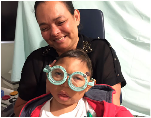
Infants with CZS, similar to other infants with brain abnormalities, and neurological visual impairment may have limited power of accommodation, poor visual acuity, high refractive errors, and strabismus44-46.
Overcorrection done to improve accommodation has been shown to result in immediate improvement in binocular vision in 62% of the children (Figure 1)44.
Since ZIKV infection is an emerging disease, a larger number of studies are required to better address this infection. Past studies have highlighted hearing loss, visual impairment, and neuromuscular and neurodevelopmental abnormalities as common long-term sequelae associated with congenital viral infections33,47.
CONCLUSIONS
ZIKV infection has recently emerged as a major global threat to pregnancies. Given the serious congenital complications that can develop due to ZIKV infection and the substantial long-term consequences, including seizures, visual and hearing impairment, feeding difficulties, and significant developmental delay, a strong and rapid global public health and research response to the virus is essential to limit and prevent the major health, social, and economic impact of the virus and to advance the development of therapeutics, vaccines, and improved diagnostics. All newborns suspected of CZS infection must undergo at least one eye examination, including a dilated fundus examination as well as auditory and global assessment. Longitudinal monitoring of the infants with possible CZS is essential for a complete characterization of the visual and systemic outcomes. The pandemic potential of the disease requires research toward the development of a vaccine and greater efforts in vector control.
REFERENCES
1. Dick GW, Kitchen SF, Haddow AJ. Vírus da zika. I. Isolamentos e especificidade serológica. Trans R Soc Trop Med Hyg. 1952;46:509-20.
2. MacNamara FN. Vírus da zika: um relatório sobre três casos de infecção humana durante uma epidemia de icterícia na Nigéria. Trans R Soc Trop Med Hyg.1954;48:139-45.
3. Musso, D, Cao-Lormeau, VM, and Gubler, DJ. Vírus da zika: seguindo o caminho da dengue e chikungunya? Lancet. 2015;386:243-4.
4. Duffy MR, Chen T-H, Hancock WT, et al. Surto do vírus da zika na ilha de Yap, Estados Federados da Micronésia. N Engl J Med. 2009;360:2536-43.
5. Cao-Lormeau VM, Roche C, Teissier A, et al. Vírus da zika, Polinésia Francesa, Pacífico Sul, 2013. Emerg Infec Dis. 2014;20:1085-6.
6. Avaliação rápida do risco: Surto de infecção pelo vírus da zika, Polinésia Francesa. Estocolmo: Centro Europeu para Prevenção e Controle da Doença, 14 de fevereiro de 2014 Disponível em: http://ecdc.europa.eu/en/publications/Publications/Zika-virus-French-Polynesia-rapid-risk-assessment.pdf Último acesso: 14 de dezembro de 2017.
7. Zanluca C, Melo VC, Mosimann AL, Santos GI, Santos CN, Luz K. Primeiro relatório de transmissão autóctone do vírus da zika no Brasil. Mem Inst Oswaldo Cruz. 2015;110:569-72.
8. Campos GS, Bandeira AC, Sardi SI. Surto do vírus da zika, Bahia, Brasil. Emerg Infec Dis. 2015;21(10):1885-6.
9. Surtos do vírus da zika nas Américas. Wkly Epidemiol Rec. 2015;90:609-610
10. Centro Europeu para Prevenção e Controle de Doenças. Avaliação rápida do risco: Epidemia do vírus da zika nas Américas: potencial associação com microcefalia e Síndrome de Guillain-Barré. 10 de Dezembro de 2015. Disponível em: https://ecdc.europa.eu/sites/portal/files/media/en/publications/Publications/zika-virus-americas-association-with-microcephaly-rapid-risk-assessment.pdf- Último acesso: 14 de dezembro de 2017.
11. Organização Mundial da Saúde (OMS). Microcefalia e Síndrome de Guillain-Barré pelo vírus da zika. Relatório da situação. 17 de março de 2016. Disponível em: http://apps.who.int/iris/bitstream/10665/204633/1/zikasitrep_17Mar2016_eng.pdf Último acesso: 14 de dezembro de 2017.
12. Possível associação entre infecção pelo vírus da zika e microcefalia — Brasil, 2015. MMWR Morb Mortal Wkly Rep. 2016;65:59-62.
13. Victora CG, Schuler-Faccini L, Matijasevich A, Ribeiro E, Pessoa A, Barros FC. Microcefalia no Brasil: como interpretar os números relatados? Lancet. 2016;387:621-4
14. Oliveira Melo AS, Malinger G, Ximenes R, Szejnfeld PO, Alves Sampaio S, Bispo de Filippis AM. Infecção intrauterina pelo vírus da zika causa anormalidade cerebral e microcefalia no feto: ponta do iceberg? Ultrasound Obstet Gynecol. 2016;47:6-7.
15. Gourinat AC, O'Connor O, Calvez E, Goarant C, Dupont-Rouzeyrol M. Detecção do vírus da zika na urina. Emerg Infec Dis. 2015;21:84-6.
16. Pinto Junior VL, Luz K, Parreira R, Ferrinho P. Vírus da zika: uma revisão para os médicos. Acta Med Port. 2015;28(6):760-5.
17. Castanha PM, Cordeiro MT, Martelli CM, Souza WV, Marques ET Jr, Braga C. Força de infecção dos serotipos da dengue em um estudo baseado na população do Nordeste do Brasil. Epidemiol Infect. 2013;141:1080-8.
18. Centro para Controle e Prevenção de Doenças. Guia de testes para infecção pelo vírus da zika para laboratórios dos E.U.A.. Atualizado em 24 de julho de 2017. Disponível em: https://www.cdc.gov/zika/laboratories/lab-guidance.html Acessado em 5 de fevereiro de 2018.
19. Centro para Controle e Prevenção da Doença. Transmissão e riscos. http://www.cdc.gov/zika/transmission/index.html. Acessado em 01 de agosto de 2016.
20. Turmel JM, Abgueguen P, Hubert B, et al. Transmissão sexual tardia do vírus da zika relacionada à persistência no sêmen. Lancet. 2016;387(10037):2501.
21. Infecção pelo vírus da zika e transplante de órgãos sólidos: um novo desafio. Am J Transplant 2016 doi: 10.1111/ajt.14047.
22. Motta IJF, Spencer BR, Cordeiro da Silva SG, et al. Evidência para transmissão do vírus da zika por transfusão de plaquetas. N Engl J Med 2016; 375:1101- 3.
23. de Araújo TVB, Rodrigues LC, de Alencar Ximenes RA, et al. Associação entre infecção pelo vírus da zika e microcefalia no Brasil de janeiro a maio de 2016: relatório preliminar de um estudo de caso com controle. Lancet Infect Dis. 2016;16(12):1356-63.
24. Miner JJ, Sene A, Richner JM, et al. Infecção pelo vírus da zika em camundongos causa panuveíte com eliminação do vírus nas lágrimas. Cell Rep 2016;16(12):3208-18.
25. Bingham AM, Cone M, Mock V, et al. Comparação dos resultados de testes para RNA do vírus da zika em amostras de urina, soro e saliva de pessoas com doença do vírus da zika associada a viagens - Flórida, 2016. MMWR Morb Mortal Wkly Rep 2016;65(18):475-8.
26. Russell K, Oliver SE, Lewis L, et al. Atualização: Orientações provisórias para a avaliação e o gerenciamento de crianças com possível infecção congênita pelo vírus da zika—Estados Unidos, agosto 2016. MMWR Morb Mortal Wkly Rep. 2016;65(33):870-8.
27. Brasil P, Pereira JP Jr, Moreira ME, Ribeiro Nogueira RM, Damasceno L, Wakimoto M, et al. Infecção pelo vírus da zika em mulheres grávidas no Rio de Janeiro. N Engl J Med. 2016;375(24):2321-34.
28. Cauchemez S, Besnard M, Bompard P, et al. Associação entre o vírus da zika e microcefalia na Polinésia Francesa, 2013-15: um estudo retrospectivo. Lancet. 2016;387(10033):2125-32.
29. Johansson MA, Mier-y-Teran-Romero L, Reefhuis J, Gilboa SM, Hills SL. Zika e o risco de microcefalia. N Engl J Med. 2016;375(1):1-4.
30. Honein MA, Dawson AL, Petersen EE, et al. Defeitos de nascença entre fetos e recém-nascidos de mulheres norte-americanas com evidência de possível infecção pelo vírus da zika durante a gestação. JAMA. 2017;317(1):59-68.
31. Rasmussen SA, Jamieson DJ, Honein MA, Peterson LR. Vírus da zika e defeitos congênitos – revisão da evidência de causalidade. N Engl J Med. 2016;374:1981-7.
32. Cugola FR, Fernandes IR, Russo FB, et al. A cepa brasileira do vírus da zika causa defeitos de nascença em modelos experimentais. Nature. 2016;534:267- 71
33. Moore CA, Staples JE, Dobyns WB, et al. Caracterizando o padrão de anomalias na síndrome congênita do vírus da zika para médicos pediatras. JAMA Pediatr. 2017;171(3):288-95.
34. Van der Linden V, Pessoa A, Dobyns W, et al. Descrição de 13 crianças nascidas no período de outubro de 2015 a janeiro de 2016 com infecção congênita pelo vírus da zika sem microcefalia ao nascimento - Brasil. MMWR Morb Mortal Wkly Rep. 2016;65:1343-8.
35. Ventura CV, Maia M, Bravo-Filho V, Góis AL, Belfort Jr R. Vírus da zika no Brasil e atrofia macular em uma criança com microcefalia. Lancet. 2016;387(10015):228.
36. Ventura CV, Maia M, Ventura BV, et al. Achados oftalmológicos em crianças com microcefalia e infecção intra-uterina presumida pelo vírus da zika. Arq Bras Oftalmol. 2016;79:1-3.
37. de Paula Freitas B, de Oliveira Dias JR, Prazeres J, et al. Achados oculares em crianças com microcefalia associada a infecção congênita presumida pelo vírus da zika em Salvador, Brasil. JAMA Ophthalmol. 2016 Feb 9. doi: 10.1001/jamaophthalmol.2016.0267.
38. Ventura CV, Maia M, Travassos SB, et al. Fatores de risco associados a achados oftalmoscópicos identificados em crianças com infecção congênita presumida pelo vírus da zika. JAMA Ophthalmol. 2016; 134(8):912-8.
39. Miranda HA II, Costa MC, Frazão MA, Simão N, Franchischini S, Moshfeghi DM. Espectro expandido de achados oculares congênitos em casos de microcefalia com infecção presumida pelo vírus da zika. Ophthalmology. 2016;123(8):1788-94.
40. Ventura LO, Ventura CV, Lawrence L, et al. Deficiência visual em crianças com síndrome congênita do vírus da zika. J AAPOS. 2017;21:295-299.e2.
41. Li C, Xu D, Ye Q, et al. O vírus da zika interrompe o desenvolvimento do progenitor neural e leva à microcefalia em camundongos. Cell Stem Cell. 2016;19:120-6.
42. Wu K, Zuo G, Li X, et al. A transmissão vertical do vírus da zika visando as células gliais radiais afeta o desenvolvimento do córtex de camundongos descendentes. Cell Res. 2016;26:645-54.
43. Van der Pol AN, Mao G, Yang Y, Ornaghi S, Davis JN. Alvos do vírus da zika no cérebro em desenvolvimento. J. Neurosci. 2017;37:2161-75.
44. Ventura LO, Lawrence L, Ventura CV, et al. Resposta à correção de erros de refração e hipoacomodação em crianças com síndrome congênita do vírus da zika. J AAPOS. 2017;21(6):480-4.
45. Jacobson L, Ygge J, Flodmark O, Ek U. Características visuais e perceptivas, mobilidade ocular e estrabismo em crianças com leucomalácia periventricular. Strabismus 2002;10:179-83.
46. Guzzetta A, Mercuri E, Cioni G. Transtornos visuais em crianças com lesões cerebrais: 2. Deficiência visual associada a paralisia cerebral. Eur J Paediatr Neurol 2001;5:115-9.
47. Leal MC, Muniz LF, Ferreira TS, et al. Perda auditiva em crianças com microcefalia e evidência de infecção congênita pelo vírus da zika - Brasil, novembro de 2015 – maio de 2016. MMWR Morb Mortal Wkly Rep. 2016:65(34):917-9.
Funding: No specific financial support was available for this study.
CEP Approval: Not applicable.
Disclosure of potential conflicts of interest: None of the authors have any potential conflict of interest to disclose.
Received on:
December 20, 2017.
Accepted on:
February 15, 2018.
 eOftalmo está licenciada com uma Licença Creative Commons Atribuição-NãoComercial 4.0 Internacional.
![]() © 2026 Todos os Direitos Reservados
© 2026 Todos os Direitos Reservados


 Ler em português
 Português PDF
 Imprimir
 Enviar este artigo por email
 Como citar este artigo
 Enviar um comentário
 Mendeley
 Pocket