

Ana Laura de Araujo Moura
DOI: 10.17545/eOftalmo/2023.0037
Este artigo pertence à Edição Especial Neuroftalmologia por imagem: acima e além
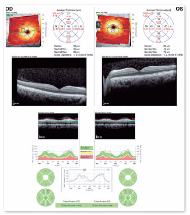
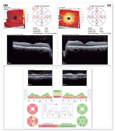
A 27-year-old man presented to the ophthalmology coherence tomography was performed immediately clinic complaining of vision loss in the right eye after after traumatic optic neuropathy (Figure 1) and after 3 an accidental fall from his height. His visual acuity months (Figure 2). The retinal nerve fiber (RNF) and was “no light perception” in the right eye and 20/20 in ganglion cell (GC) layers were initially normal, despithe left eye. Ocular motility, biomicroscopy, and fun-te the extreme visual acuity loss in the right eye. At doscopy findings were unremarkable, and there was the time, the most reliable information about optic an afferent pupillary defect in the right eye. Optical nerve integrity was obtained from the pupillary light reflex. Three months later, there was a severe decrease in the thicknesses of both the RNF and GC layers.
The optic nerves are vulnerable to direct and indirect trauma. Direct injury can be caused by penetrating trauma, such as that associated with orbital fractures or foreign bodies. Indirect injury, which is the most common injury type, occurs due to the forces generated by head trauma and transmitted to the optic nerve via the orbital apex and optic canal. In such a case, the head trauma can be light (to the malar or frontal areas) or severe and can occur along with traumatic brain injury1.
The clinical findings include reduced visual acuity, visual field defects, color vision loss and afferent pupillary defects. Usually, in indirect optic neuropathy, the optic nerve appears normal on fundoscopy during the initial weeks after a trauma but becomes atrophic subsequently2.
Despite attempts to use steroids and perform surgery, the treatment for traumatic optic neuropathy remains controversial because of the lack of evidence-based guidelines3.
REFERENCES
1. Warner N, Eggenberger E. Traumatic optic neuropathy: a review of the current literature. Curr Opin Ophthalmol. 2010;21(6):459-62.
2. Cunha LP, Costa-Cunha LVF, Malta RFS, Monteiro MLR. Comparison between retinal nerve fiber layer and macular thickness measured with OCT detecting progressive axonal loss following traumatic optic neuropathy. Arq Bras Oftalmol. 2009:72(5):622-5.
3. Chaon BC, Lee MS. Is there treatment for traumatic optic neuropathy? Curr Opin Ophthalmol. 2015;26(6):445-9.
AUTHOR INFORMATION

Funding: No specific financial support was available for this study.
Conflict of interest: None of the authors have any potential conflict of interest to disclose.
Received on:
June 7, 2023.
Accepted on:
June 12, 2023.
 eOftalmo está licenciada com uma Licença Creative Commons Atribuição-NãoComercial 4.0 Internacional.
![]() © 2026 Todos os Direitos Reservados
© 2026 Todos os Direitos Reservados
 Ler em português
 Português PDF
 Imprimir
 Enviar este artigo por email
 Como citar este artigo
 Enviar um comentário
 Mendeley
 Pocket