

Marina Gonçalves Vieira; Daniella Pedra Vasconcellos; Erika Marques Demori
DOI: 10.17545/eOftalmo/2023.0033
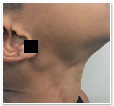
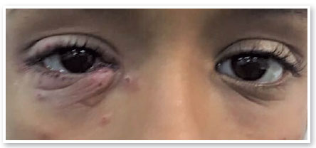
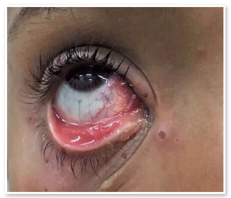
Parinaud oculoglandular syndrome is a rare condition characterized by the presence of granulomatous conjunctivitis associated with ipsilateral lymphadenopathy1, the most frequent cause of which is Bartonella henselae infection. However, other etiologies such as herpes, blastomycosis, tuberculosis, syphilis, and, less frequently, sporotrichosis, are also described1,2. Sporotrichosis is a subacute to chronic mycosis1 caused by contact with contaminated soil or sick animals and inhalation of spores2. We report a case of this syndrome secondary to infection by the fungus Sporothrix schenckii, which characteristically manifests with subcutaneous nodules along the path of the lymphatic vessels. Ocular involvement occurs in different ways, from dacryocystitis and conjunctivitis to severe cases of choroiditis and endophthalmitis3,4. A 10-year-old male patient from Rio de Janeiro sought care complaining of red eye and presenting eyelid lesions with onset 3 days earlier. Ectoscopy showed multiple round lesions on the eyelids of the right eye, associated with submandibular and ipsilateral cervical lymph node enlargement (Figure 1). Visual acuity was 20/20 inboth eyes. He presented conjunctival hyperemia and granulomatous follicular reaction in the inferior tarsal conjunctiva of the right eye (Figures 2 and 3). The examination of the left eye by tonometry and fundoscopy showed no alterations. Complementary tests were requested for further investigation, including serology for syphilis and HIV, chest X-ray, and tuberculin test. In addition, conjunctival material was collected and sent for culture. Sporothrix sp. growth was obtained in Sabouraud agar and Mycosel agar, thus confirming the diagnostic suspicion of sporotrichosis. Moreover, the patient reported contact with cats, which were later also diagnosed and treated for the disease. Itraconazole was the therapy of choice, at a dose of 100mg twice a day, for a period of 90 days, and the condition resolved in the 1st month of treatment. The state of Rio de Janeiro is experiencing an epidemic of the disease, where it is no longer infrequent. Early diagnosis and correct treatment by an ophthalmologist are of utmost importance to avoid loss of visual function.
REFERENCES
1. Ribeiro ASA, Bisol T, Menezes MS. Parinaud’s oculoglandular syndrome caused by Sporotrichosis. Rev Bras Oftalmol. 2010;69 (5):317-22.
2. Madureira LS, Gatti RF, Prohmann CM, Sanmiguel J, Almeida MTG, Mattar FRO, et al. Parinaud’s Oculoglandular Syndrome Caused by Sporothrix schenckii. SPDV. 2018;76(4):429-433.
3. Yamagata JPM, Rudolph FB, Nobre MCL, Nascimento LV, Sampaio FMS, Arinelli A, et al. Ocular sporotrichosis: A frequently misdiagnosed cause of granulomatous conjunctivitis in epidemic areas. Am J Ophthalmol Case Rep. 2017 Dec;8:35-38.
4. Ribeiro CR, Silva BP, Costa ADAA, Basile A Neto, Vieira LA, Lima MA, et al. Ocular Sporotrichosis. Am J Ophthalmol Case Rep. 2020 Auh 6;19:100865.
5. Furtado LO, Biancardi AL, Cravo LMS, Anjo RPP, Moraes HV Junior. Ocular sporotrichosis: atypical manifestations. Rev Bras Oftalmol. 2019;78(1):59-61.
AUTHORS INFORMATIONS


Funding: No specific financial support was available for this study.
Conflict of interest: None of the authors have any potential conflict of interest to disclose.
Received on:
November 24, 2022.
Accepted on:
July 2, 2023.
 eOftalmo está licenciada com uma Licença Creative Commons Atribuição-NãoComercial 4.0 Internacional.
![]() © 2026 Todos os Direitos Reservados
© 2026 Todos os Direitos Reservados

 Ler em português
 Português PDF
 Imprimir
 Enviar este artigo por email
 Como citar este artigo
 Enviar um comentário
 Mendeley
 Pocket