

Juliana Prazeres
DOI: 10.17545/eOftalmo/2023.0016
Este artigo pertence à Edição Especial Dominando a arte da cirurgia vitreorretiniana: técnicas e dicas
Proliferative vitreoretinopathy (PVR) can occur as a complication of rhegmatogenous retinal detachment (RD) and can have poor visual outcomes. A specific manifestation of PVR is the formation of subretinal bands, which is associated with an irregular scarring process where membranes develop beneath the detached retina. This condition arises owing to the abnormal growth of retinal cells, which leads to the development of tractional bands that can contribute to recurrent RD1.
Although there is a connection between recurrent rhegmatogenous RD and PVR, it is not obligatory to treat subretinal fibrosis. In certain cases, particularly those in which patients experience chronic RD complicated by subretinal PVR, anatomical success can be achieved through scleral buckling surgery without the requirement for posterior vitrectomy to treat subretinal fibrosis2,3.
We present a case of a 37-year-old patient with high myopia, who complained of decreased visual acuity in the left eye for the past 7 months. Ophthalmic examination revealed a visual acuity of 20/200. Fundoscopic examination showed subtotal RD, with the involvement of the inferior, nasal, and temporal retina, as well as the presence of extensive subretinal fibrosis bands, indicating PVR stage C. Inferior lattice degeneration was observed, and no retinal tears were identified.
Scleral buckling was selected to treat the RD. Chandelier-assisted scleral buckling, which uses a wide-angle system, has demonstrated effectiveness and favorable outcomes in the treatment of rhegmatogenous RD3.
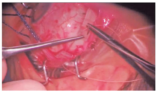
After performing a 360° peritomy, the extraocular muscles are isolated using appropriate hooks and 2-0 cotton. Subsequently, sclerotomy is performed for the insertion of the chandelier. Using a noncontact wide-angle visualization system, cryotherapy is performed in the region of inferior lattice degeneration where microholes are present. After passing band 42 under the rectus muscles, the distance from the limbus to the area with retinal microholes is measured, and the band is sutured using 5-0 polyester suture thread. When employing this technique, it is important to posteriorize the suture knot to minimize the risk of suture extrusion. After delaminating the sclera using blade no. 11 and cauterizing the choroidal tissue, drainage of subretinal fluid is performed using the suture thread needle. As seen in the video, the drained fluid is thick and its quantity is small, which is indicative of a chronic RD. A 6-cm portion of the band is then cut and sutured.
There was progressive absorption of the subretinal fluid, as evidenced by retinography and optical coherence tomography examinations.
REFERENCES
1. Nemet A, Moshiri A, You G, Loewenstein A, Moisseiev E.A Review of Innovations in Rhegmatogenous Retinal Detachment Surgical Techniques. J Ophthalmol. 2017;2017:4310643.
2. Yao Y, Jiang L, Wang Z, Zhang M. Scleral buckling procedures for longstanding or chronic rhegmatogenous retinal detachment with subretinal proliferation. Ophthalmology. 2006;113(5):821-5.
3. Roca JA, Maia M, Cruz NFS, Polizelli MU, Chhablani J, Gangakhedkar S, et al. Non-contact wide-angled visualization with chandelier-assisted scleral buckling for primary uncomplicated rhegmatogenous retinal detachment. Graefes Arch Clin Exp Ophthalmol. 2020;258(9):1857-1861.
AUTHOR INFORMATION
Funding: No specific financial support was available for this study.
Conflict of interest: None of the authors have any potential conflict of interest to disclose.
Received on:
July 9, 2023.
Accepted on:
July 12, 2023.
 eOftalmo está licenciada com uma Licença Creative Commons Atribuição-NãoComercial 4.0 Internacional.
![]() © 2026 Todos os Direitos Reservados
© 2026 Todos os Direitos Reservados

 Ler em português
 Português PDF
 MP4
MP4
 Imprimir
 Enviar este artigo por email
 Como citar este artigo
 Enviar um comentário
 Mendeley
 Pocket