

Carlos Augusto Moreira Neto1; Gabriel Martins Duda2
DOI: 10.17545/eOftalmo/2023.0007
Subretinal fibrosis (SRF) is a nonspecific process resulting from damage to the retinal pigment epithelium, retina, and underlying choroid. SRF is defined as a whitish material, which can be pigmented and of coarse appearance, in the subretinal space1,2.
Cytokines, immunoglobulins, and T lymphocytes are believed to generate fibrosis by interacting with retinal pigmented epithelial (RPE) cells, Muller’s cells, and choroidal fibrocytes. However, the nature and its implication may vary in different eye diseases3.
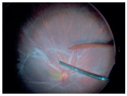
The video shows a 31-year-old male patient with visual acuity of light perception. He has previously undergone two corneal transplants and presented cataracts and retinal detachment.
The patient was submitted to lens phacoemulsification with intraocular lens implantation and 25-gauge pars plana vitrectomy, using Chandelier accessory illumination and scleral indentation to remove the vitreous base.
The patient presented intense SRF in posterior pole and optic disc, and retinotomies were performed to facilitate subretinal fibrosis removal. Optical fibers were also used to help remove the membranes.
After releasing temporal retina of traction, peridiscal SRF and nasal retina were removed.
Air-fluid exchange was performed several times in order to visualize traction forces and guide the removal of fibroses that were still present.
Even with subretinal membrane removal, the air in the vitreous cavity showed nasal retina traction. Thus, micheal peek and 25-gauge clamp claw were used to provide greater force in clamping and total removal of SRF.
After total removal of the tractions, the retina is then completely attached.
Then, photocoagulation is performed in 360 degree retinotomies. At the end, silicone oil is injected.
REFERENCES
1. Wallyn RH, Hilton GF. Subretinal fibrosis in retinal detachment. Arch Ophthalmol. 1979;97(11):2128-9.
2. Golzarri MF, Cheja-Kalb R, Concha-Del-Río LE, Gonzalez-Salinas R, Arellanes-García L. Risk factors for subretinal fibrosis in patients with Vogt Koyanagi Harada syndrome. Ocul Immunol Inflamm. 2022;30(2):265-9.
3. Lertsumitkul S, Whitcup SM, Nussenblatt RB, Chan CC. Subretinal fibrosis and choroidal neovascularization in Vogt-Koyanagi-Harada syndrome. Graefes Arch Clin Exp Ophthalmol. 1999;237(12):1039-45.
AUTHOR’S INFORMATION

Financiamento: No specific financial support was available for this study.
Conflitos de Interesse: None of the authors have any potential conflict of interest to disclose.
Received on:
January 29, 2023.
Accepted on:
March 1, 2023.
 eOftalmo está licenciada com uma Licença Creative Commons Atribuição-NãoComercial 4.0 Internacional.
![]() © 2026 Todos os Direitos Reservados
© 2026 Todos os Direitos Reservados

 Ler em português
 Português PDF
 MP4
MP4
 Imprimir
 Enviar este artigo por email
 Como citar este artigo
 Enviar um comentário
 Mendeley
 Pocket