

Juliana Angélica Estevão de Oliveira1; Natália Lopes Leal2; Mariluze Maria Souza Sardinha2,3
DOI: 10.17545/eOftalmo/2022.0016
ABSTRACT
Fraser’s Syndrome is a rare autosomal recessive disease whose main ocular manifestation is cryptophthalmos. The abortive variant manifests as coloboma of the medial upper eyelid, abnormal upper fornix, and symblepharon. This article reports two cases of FS in female children, who underwent upper fornix and upper eyelid reconstruction due to abortive cryptophthalmos in eyes with visual potential. The most important difficulty was the lack of tissue available for reconstruction, as one patient was only 33 days old and the other less than 3 years old. In both cases, there was recurrence of the symblepharon. In the first one, the oral mucosa was not enough to cover the eyeball, and amniotic membrane and conformator were not available. In the second, the presence of conjunctival abnormalities was not recognized intraoperatively. Further surgeries will be delayed until patients have more tissue laxity since both corneas are not exposed.
Keywords: Fraser syndrome; Coloboma, Conjunctiva; Myocutaneous flap; Syndactyly.
RESUMO
A Síndrome de Fraser é uma doença autossômica recessiva rara, cuja principal manifestação ocular é a criptoftalmia. A variante abortiva se manifesta como coloboma da pálpebra superior medial, fórnice superior anormal e simbléfaro. Este artigo relata dois casos de SF em crianças do sexo feminino, submetidas a reconstrução de fórnice e pálpebra superior por criptoftalmia abortiva em olhos com potencial visual. A dificuldade mais importante foi a falta de tecido disponível para reconstrução, já que uma paciente tinha apenas 33 dias e a outra menos de 3 anos. Em ambos os casos, houve recorrência do simbléfaro. No primeiro, a mucosa oral não foi suficiente para cobrir o globo ocular e, membrana amniótica e conformador não estavam disponíveis. No segundo, a presença de anormalidades conjuntivais não foi reconhecida no intraoperatório. Outras cirurgias serão adiadas até que os pacientes tenham mais frouxidão tecidual, uma vez que ambas as córneas não estão expostas.
Palavras-chave: Síndrome de Fraser; Coloboma; Conjuntiva; Retalho miocutâneo; Sindactilia
INTRODUCTION
Fraser’s Syndrome (FS) is a rare, autosomal recessive disease, whose diagnosis is based on phenotypic features1,2. The prevalence rate of consanguinity in families with FS is estimated at 27%3. Ocular anomalies occur in up to 83% of patients and manifest most commonly as cryptophthalmos1. Cryptophthalmos can be classified into three types: complete, incomplete, and abortive4. The complete form is characterized by continuity of the skin over a disorganized ocular structure4,5. In the incomplete form, lateral eyelid structures are present. In the abortive variant, there is coloboma of the medial upper eyelid, abnormal superior fornix and symblepharon5. In this article, we report two cases of this rare syndrome to help its recognition and to discuss difficulties of its ophthalmological rehabilitation.
CASES REPORT
Case 1
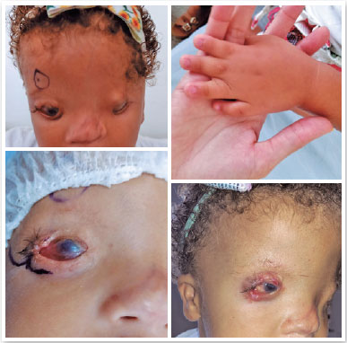
A 33-month-old female patient diagnosed with FS had bilateral abortive cryptophthalmos, hypertelorism, widely spaced eyebrows, low hair implantation on forehead, cleavage along the midplane of the nose, low ear implantation, syndactyly in the central three fingers of hands, umbilical hernia, single kidney, and abnormalities of external genitalia. There was a family history of parental consanguinity and an affected sibling. Ophthalmological examination showed ability to pursuit objects in right eye (OD), to follow a light source in left eye (OS) and, in both eyes (OU), keratinized cornea preventing visualization of the anterior chamber, total limbic insufficiency, shallow lower fornix, coloboma of upper eyelids and symblepharon. Right upper eyelid reconstruction was planned. Dissection of corneal adhesions was performed, and an oral mucous graft was sutured as the posterior lamella of the rudimentary upper eyelid. The sclera was left bare due to tissue unavailability, as well as symblepharon ring not available. A full-thickness lower eyelid graft, in pentagon format, was sutured to upper eyelid defect. Primary closure was chosen for the lower eyelid defect. Approximately 30 days later, recurrence of the symblepharon was observed (Figure 1).
Case 2
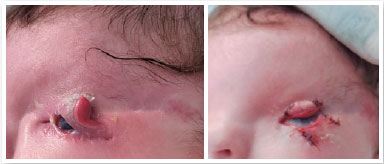
A 33-days-old female patient diagnosed with FS had abortive cryptophthalmos in OS and complete in OD, syndactyly in the four medial fingers of hands, deformity and low ear implantation, low hair implantation on forehead, tracheal stenosis, and abnormality of the external genitalia. Both cases might be distant relatives since their families come from the same region. Ophthalmological examination showed ability to follow a light source in OS, coloboma of the upper eyelid, symblepharon, keratinized cornea preventing visualization of the anterior chamber and shallow lower fornix. Cranial computed tomography (CT) scan revealed microphthalmia and absence of lens in OD, enlarged eyeball in OS. Ocular ultrasound showed small optical vesicles and absence of the lens in OD, 21.50 mm of axial length and evident cupping in OS. It was decided to perform reconstruction of the left upper eyelid first and then trabeculotomy. Dissection of corneal adhesions was performed and there was normal looking conjunctiva in the fornix and posterior lamella. Thus, no mucosal graft was needed. A flap of full thickness of the lower eyelid was sutured to the upper eyelid (Mustardé flap). Second intention healing was chosen for the lower eyelid defect. About ten days later, partial dehiscence and recurrence of the symblepharon were noticed. Subsequent procedures included new flap suture and bulbar conjunctival graft transplantation to superior fornix. After five weeks, division of the flap was performed, and the lower eyelid was primary closed after lateral canthotomy to released tension (Figure 2).
DISCUSSION
Abortive cryptophthalmos is potentially vision-threatening because of corneal exposure resulting from upper eyelid coloboma and symblepharon that limits motility of the globe4. This is the main indication for early surgery in eyes with visual potential and it was present in both reported cases2,4. The purpose of surgery is to reconstruct the upper eyelid and superior fornix4. Orbital CT and ocular color doppler imaging can be used prior to surgery to help evaluate ocular globe integrity5. Multiple techniques have been described, but most surgeons prefer eyelid-sharing techniques like Cutler–Beard or a full-thickness lower eyelid Mustardé-type switch flap with additional posterior lamellar reconstruction, as performed in the second reported case1,2,4. The challenge is often compounded by the lack of tissue laxity in children, the risk of inducing iatrogenic amblyopia due to occlusion of the visual axis and the risk of scar retraction and distortion of the donor lower eyelid4.
Various materials have been used to reconstruct the fornix, including oral mucous membrane, hard palate, conjunctiva, scleral grafts and amniotic membrane1,4,5. It is reported that amniotic membrane was superior to buccal mucous membrane and hard palate in the maintenance of the fornix in cryptophthalmic repair4,5. Its major advantage is the ability to reduce inflammation and scarring while promoting epithelization4.
Recurrence of symblepharon is comparatively common after surgery in abortive cryptophthalmos and tend to recur in the same area where they were observed at the first presentation4. Shell conformers or scleral lenses can be helpful to maintain conjunctival sac and avoid new corneopalpebral adhesions5.
The normal looking conjunctiva of the upper fornix of the second reported case was not healthy and a bulbar conjunctival graft was subsequently needed. There was recurrence of the symblepharon on both cases, a risk that could have been reduced if amniotic membrane or a symblefharon ring had been available.
Although surgical reconstruction to improve visual acuity up to 20/100 has been reported, visual prognosis is typically poor1,2. These issues must be discussed carefully with the parents before embarking on surgical treatment, which will be different in each case, depending on the extent of the ocular abnormality and the visual potential2,4. Oculoplastic surgical rehabilitation in Fraser syndrome is extremely challenging, and frequently multiple operations will be required for these patients due to degree of scarring, contracture, and the health of the cornea1,2,4,5. Generally, reconstruction of the eyelids should precede any procedures to the cornea and the priority is rehabilitating the eye with visual potential2.
The reported cases can help in the clinical diagnosis of this rare syndrome. Furthermore, they show that the major challenge of eyelid reconstruction in these cases is limited tissue laxity. The use of amniotic membrane, when available, can help prevent symblepharon recurrence.
REFERENCES
1. Tran AQ, Lee BW, Alameddine RM, Korn BS, Kikkawa DO. Reconstruction of Unilateral Incomplete Cryptophthalmos in Fraser Syndrome. Ophthalmic Plast Reconstr Surg. 2017;33 (3S Suppl 1):S73-S75.
2. Saleh GM, Hussain B, Verity DH, Collin JR. A surgical strategy for the correction of Fraser syndrome cryptophthalmos. Ophthalmology. 2009;116(9):1707-1712.e1.
3. Mbonda A, Endomba FT, Kanmounye US, Nkeck JR, Tochie JN. Diagnosis of Fraser syndrome missed out until the age of six months old in a low-resource setting: a case report. BMC Pediatr. 2019;19(1):292.
4. Ding J, Hou Z, Li Y, Lu N, Li D. Eyelid and fornix reconstruction in abortive cryptophthalmos: a single-center experience over 12 years. Eye (Lond). 2017;31(11):1576-1581.
5. Liu Z, Xie B, Li Y, Ding J, Li D. Reconstruction strategy in isolated complete Cryptophthalmos: a case series. BMC Ophthalmol. 2019;19(1):165.
AUTHOR’S INFORMATION
Funding: No specific financial support was available for this study
Approved by the following research ethics committee: Hospital Professor Edgard Santos (CAEE: 50133521.1.0000.0049).
Conflict of interest: None of the authors have any potential conflict of interest to disclose
Received on:
August 26, 2022.
Accepted on:
October 11, 2022.
 eOftalmo está licenciada com uma Licença Creative Commons Atribuição-NãoComercial 4.0 Internacional.
![]() © 2026 Todos os Direitos Reservados
© 2026 Todos os Direitos Reservados

 Ler em português
 Português PDF
 Imprimir
 Enviar este artigo por email
 Como citar este artigo
 Enviar um comentário
 Mendeley
 Pocket