

Silvana V. Lazary1; Fernanda T. Krieger2
DOI: 10.17545/eOftalmo/2019.0025
ABSTRACT
Strabismus and diplopia are rare complications that can occur after refractive surgery. In this report, we describe the case of a male patient who underwent refractive surgery (LASIK) using the monovision technique and developed strabismus and persistent diplopia two years later even after monovision correction with new refractive surgery, requiring strabismus surgery.
Keywords: Diplopia; Strabismus; LASIK.
RESUMO
O estrabismo e a diplopia são complicações raras, mas possíveis após uma cirurgia refrativa. Nesse trabalho descrevemos o caso de um paciente do sexo masculino submetido à cirurgia refrativa (LASIK) usando a técnica de monovisão e que dois anos após desenvolveu estrabismo e diplopia persistente mesmo após a correção da monovisão com nova cirurgia refrativa, sendo necessária cirurgia de estrabismo.
Palavras-chave: Diplopia; Estrabismo; LASIK.
RESUMEN
El estrabismo y la diplopía son complicaciones raras, pero posibles tras una cirugía refractiva. En este trabajo, describimos el caso de un paciente del sexo masculino sometido a la cirugía refractiva (LASIK) usando la técnica de monovisión y que dos años después desarrolló estrabismo y diplopía persistente, aún tras la corrección de la monovisión mediante una nueva cirugía refractiva, siendo necesaria la cirugía de estrabismo.
Palabras-clave: Diplopía; Estrabismo; LASIK.
INTRODUCTION
Worldwide, refractive surgery is increasingly being used as an alternative to eyeglassesand contact lenses. Complications such as ectasia, infection, and corneal decompensation are widely known by physicians and the population.Conversely, diplopia and strabismus resulting from refractive surgery are less known potential complications with a significant negative impact on patients' lives. Repercussions in personal and professional lives are devastating and definitely affect the doctor–patient relationship and society's perception of ophthalmologists in an unfavorable manner.
Diplopia may be temporary, but it is persistent in most cases and requireseither another refractive procedure to improve the patient's condition or surgical correction of strabismus. It is important to emphasizethat some cases of diplopia are untreatable, although this is rare. The duration of onset of diplopia after refractive surgery may range from a few days to a few years.
OBJECTIVE
To report a case of diplopia and strabismus of a patient who underwent monovision refractive surgery.
CASE DETAILS
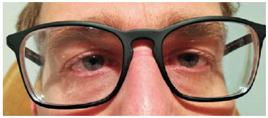
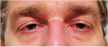
A 45-year-old male patient complainedof diplopia and strabismus for three years.The patient underwent refractive surgery for correction of myopia fifteen years ago (-3.00SPH in both eyes) and surgical retouching five years ago (Figures 1 and 2).
• Current eyeglasses: 9 temporal prism correction OU.
• Refraction: OD: -2.75D SPH 1.0.
• OS: +0.75D SPH-0.75D CYL × 120º 1.0.
• ET prefers OS.
• Prism cover test without eyeglasses: OD fixation= OE fixation: ET 25 R/L 4Δ. Same results for near fixation.
• Same measurements in all positions of gaze, including head tilting to the right and left.
• These measurements remain the same with monovision correction.
• Fusion with objective measurement.
• Conduct: Monovision correction.
• Underwent LASIK OD.
New assessment approximately six months after refractive surgery:
• No improvement in deviation and diplopia.
• VA OU 1.0 without correction.
• OD refraction: -0.25D × 90º 1.0.
• OS: +0.75 -0.75 × 180º 1.0.
• Far CrP measurement: ET 20 D/E4Δ.
• Near CrP measurement: ET' 20Δ.
• Conduct: Strabismus surgery.
• 4.0mm right medial rectus retraction/4.5mm right lateral rectus resection.
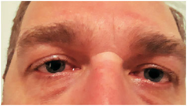
• One year after surgery.
• Far and near orthophoria.
• Titmus: 50 arc seconds (Figure 3).
DISCUSSION
Several factors may cause oculomotor system imbalance after refractive surgery. These patients usually have had previous binocular vision disorders, and refractive surgery decompensates a previously compensated condition.The main causes of decompensation are as follows: unwanted refraction correction, prismatic effect owing to decentered ablation, increased accommodative demand, anisometropia, elimination of suppression, and change of ocular dominance1.
In a study conducted including 28 patients who developed diplopia after refractive surgery, the authors reported the presence of these factors and emphasized the importance of examining these patients systematically before the surgical procedure to look for signs of ocular motricity disorders, such as eyeglasses with prism correction, which are quite often unknown to surgeons and patients2.
Development of diplopia after refractive surgery is rare (some studies estimate it to be 0.12%), and this is often not previously discussed with the patient3.More importantly, ocular motricity examination is frequently overlooked or neglected by the refractive surgeon. Thus, there may be legal consequences of not making everything clear to the patient and not performing examinations that could contraindicate or show an increased risk for complications.
In the present case, the patient had no history of strabismus, and his binocular vision after surgical monovision and strabismus correction was normal. These data suggest that a prolonged period of monovision of this magnitude was the cause of oculomotor imbalance. The degree of induced anisometropia is directly related to the harmful effects on binocular vision.A study found that patients with a difference of <1.50D between both eyes presented a 100-arc-second stereopsis, whereas patients with a difference of ≥1.50D showed 150-arc-second stereopsis4.
The onset of strabismus in this patient was noted two years after the initiation of monovision. In the literature, this period ranges from three months to two years, and some patients have recovered fusion by simply eliminating monovision in a new refractive surgery, whereas others had to undergo surgical correction of strabismus to restore binocularity. One patient was unable to recover fusion even with the ocular alignment obtained via strabismus surgery5.
Because the condition persisted for six months after the resolution of monovision, surgical correction was chosen. Some authors recommend this procedure four months after monovision correctionif strabismus does not improve5.
Considering all the factors involved in the onset of diplopia after refractive surgery is performed, all patients eligible for this surgery should have their oculomotor system assessed. After this evaluation, the patient can be classified to be at a low, medium, or high risk of diplopia and strabismus, as stated in another study2, and measures to minimize the impact of refractive surgery on the oculomotor system can be taken.
The authors of the aforementioned study proposed the following classification system:
• Low risk: Presence of myopia (<4.00D SPH anisometropia), no history of strabismus or diplopia, no history of prism correction lens use, no heterophoria upon cover test, and difference between used refraction and cycloplegic refraction of ≤0.50D SPH.
• Medium risk: Presence of hyperopia, monovision planning, and hypercorrected myopia.
• High risk: History of strabismus, presence of hyperopia with low fusional capacity, and hypo- or hypercorrected hyperopia.
Some preventive measures to prevent this complication are as follows: adjusting the correction before surgery, not letting the dominant eye be at a functional disadvantage, avoiding myopia hypercorrection and hyperopia undercorrection, evaluating fusional capacity in certain patients, and testing monovision with contact lenses in patients at risk. This monovision test does not guarantee that oculomotor system imbalance will not occur. As reported here and in studies by other authors, the onset of diplopia in these patients may take years5. Another hypothesis to be considered in our case is that the patient had a compensated low fusional amplitude prior to monovision, which was associated with anisometropia of such magnitude for an extended period and was responsible for the oculomotor system disorder.
To avoid binocular vision imbalance, another study suggests not changing the natural dominance and when operating on each eye at a different time, always operating on the dominant eye first6.
Although it is not possible to eliminate the possibility of occurrence of diplopia after refractive surgery, it is of paramount importance to consider the occurrence of this complication, to carefully examine patients for ocular motricity conditions, and to talk to them and provide advice to each patient appropriately. Thus, the potentially devastating impact on these patients' lives can be avoided.
REFERENCES
1. Gómez de Liaño-Sánchez R, Borrego-Hernando R, Franco-Iglesias G, Gómez de Liaño-Sánchez P, Arias-Puente A. Strabismus and diplopia after refractive surgery. Arch Soc Esp Oftalmol. 2012 Nov;87(11):363-7.
2. Kushner BJ, Kowall L. Diplopia after refractive surgery: occurrence and prevention. Arch Ophthalmol. 2003 Mar;121(3):315-21.
3. Minnal VR, Rosemberg JB. Refractive surgery: a treatment for a cause of strabismus. Curr Opin Ophthalmol. 2001 Jul;22(4):225-5.
4. Fawcett SL, Herman WK, Alfieri CD, Castleberry KA, Parks MP, Birch EE. Stereoacuity and foveal fusion in adults with long-standing surgical monovision. J AAPOS. 2001 Dec;5(6):342-7.
5. Pollard ZF, Greenberg MF, Bordenca M, Elliott J, Hsu V. Strabismus precipited by monovision. Am J Ophthalmol. 2011 Sep;152(3):479-482.e1.
6. Cunha RNP, Isoldi IE, Cunha M. Alterações motoras após cirurgia refrativa no paciente estrábico. Arq Bras Oftalmol. 2004;67(3):507-508.
Funding: No specific financial support was available for this study.
Disclosure of potential conflicts of interest: None of the authors have any potential conflict of interest to disclose.
Received on:
May 27, 2019.
Accepted on:
July 10, 2019.
 eOftalmo está licenciada com uma Licença Creative Commons Atribuição-NãoComercial 4.0 Internacional.
![]() © 2026 Todos os Direitos Reservados
© 2026 Todos os Direitos Reservados


 Ler em português
 Português PDF
 Imprimir
 Enviar este artigo por email
 Como citar este artigo
 Enviar um comentário
 Mendeley
 Pocket